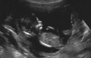
Ultrasounds

On average, we perform at least four ultrasounds per pregnancy. We will perform them at the practice – one at your first exam (at about 8 weeks) and at 12 weeks, to determine if the child has a heartbeat, whether it’s a single baby or multiple babies. Additionally, it’s important to us to establish a reliable due date.

At 20 weeks pregnancy, we will perform an elaborate ultrasound at Keik. During this specialized exam, your child is examined for a number of abnormalities. However, a positive outcome of the ultrasound does not guarantee that your baby is healthy. Small or difficult to detect abnormalities, such as, for example, a specific heart defect or autism, are not detected on an ultrasound.

We will perform another ultrasound to determine growth at 28 weeks of pregnancy. It is sometimes necessary to perform additional ultrasounds.

All echoes are and always will be snapshots: unfortunately, things can always be missed.

Early in pregnancy (before 10 weeks of pregancy) the uterus is still small and hidden behind the pubic bone. That’s why this early medical ultrasound will be performed using a small scan probe vaginally. Most women experience this without any pain. The imaging in this early term is better using an vaginal scan. At an ultrasound with 6 weeks you can only see a heartbeat at the ultrasound, because the embryo is not yet recognisable as a baby very early in pregnancy.

From a medical point of view we won’t listen to the baby’s heartbeat before 10 weeks, after 10 weeks we do.

For a vaginal ultrasound a full bladder can be in the way. That’s why we ask you to pie before you have the ultrasound.

A dating ultrasound is done to determine how far a long your pregnancy is. The term of pregnancy is best to be set between 10 and 13 weeks of pregnancy.

Till about 14 weeks the development and grow of every foetus is about the same. The sonographer can determine the age of the foetus by measuring the baby is this term of pregnancy. This way she can set your due date.

If you see a good beating hearth and a well developing foetus, the chances of a miscarriage go down with 10% to only 1-3% (depending on the duration of pregnancy). A comforting thought!

The 20 weeks ultrasound is a structural ultrasound examination. Here you can read more information about this ultrasound.

Screening

  • We will check the baby for signs of physical abnormalities.
  • At the 20 weeks ultrasound we are not able to see all abnormalities. For all ultrasounds it’s known that not all the time all abnormalities are found. Furthermore not all congenital abnormalities are present during pregnancy. If we see no abnormalities this doesn’t give guaranties for a healthy baby.
  • If we see abnormalities, the consequences for the baby aren’t always (immediately) clear. We will refer you to a specialized gynaecologist which can confirm the abnormality. But this follow-up exam can also show that there is nothing wrong with your baby, or that it is still unclear what is going on and what the impact will be.
  • Your health insurance will reimburse the 20 week ultrasound from your basic insurance.

Indication of an abnormality

If we see an indiciation of an abnormality at the 20 week ultrasound, the consequences for the baby aren’t always clear. Most of the time follow-up diagnostic testing is needed. During a consultation with the sonographer, midwife or gynaecologist you will be extensively informed about follow-up diagnostic testing. They can also answer your questions and support you. For any follow-up tests it is your decision whether you want to have these tests.

Follow-up testing

The first follow-up test will be an extended ultrasound examination, called a GUO (geavanceerd echoscopisch onderzoek). This is done by a gynaecologist at the hospital.

During this ultrasound the baby’s head, waist circumference and leg length will be measured. This way it will get clear if the baby is growing as expected. We can repeat this growth check multiple times, to get a good view of the growth of the baby: a growth curve. Also the amount of amniotic fluid will be determined.

A growth ultrasound can be performed between 22 and 40 weeks of pregnancy.

A placenta localisation is performed if during the 20 weeks ultrasound, or in some cases during a later ultrasound, the placenta is seen to be near to or covering the cervix. The ultrasound will be performed around 32 weeks of pregnancy. During this ultrasound we check if the placenta is still low in the uterus, near to the cervix, or if the placenta has moved up naturally with the growth of the uterus. A low lying placenta can be a possible hinder for a natural delivery.

We start off with an external ultrasound to measure the growth of the baby and the amount of amniotic fluid. We can also see where the placenta is located, but most of the time not exactly how low the placenta lies.

To perform the placenta localisation properly an internal ultrasound can be needed. This way we can do an accurate measurement of how far the placenta is located from the cervix. This internal exam mostly lasts a few minutes.

We will schedule a positioning ultrasound when there is a suspicion of the baby lying in a breech position around 35 weeks of pregnancy.

The sonographer can see the baby’s position with the ultrasound. Most babies lay head down, but a small number of babies (3-4%) is in a breech position. Another possibility is that the baby is in a transverse position. This is rare.

When the baby is in a breech or transverse position, we’ll look for a cause, but most of the time there is now cause for this position. Also we will check the amount of amniotic fluid and the growth of the baby.